
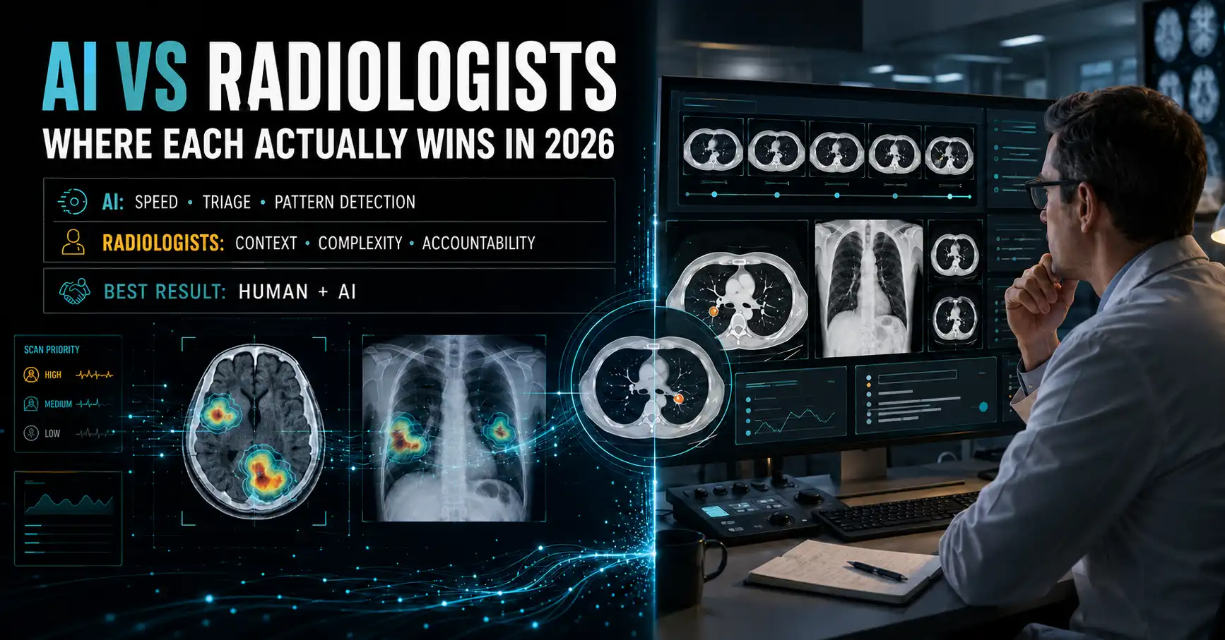
Ask a radiologist whether software is coming for the job, and you’ll usually get a tired smile. They’ve been hearing it since 2016. Ask a patient sitting on a three-week wait for a scan report, and the same question lands very differently. That is the AI vs radiologists argument in one sentence.
That gap is why the debate gets argued so badly. People treat it as one contest with one winner. The evidence splits by task instead. On some scans the software finds cancers that human readers miss. On others it cries wolf five times out of ten.
So let’s score it properly. Modality by modality, using published clinical numbers rather than vendor slides.
What “better” even means on a scan
Most AI vs radiologists comparisons collapse the moment you ask what was measured. Four things matter, and they move independently.
- Sensitivity: of the patients who have the disease, how many does the reader catch?
- Specificity and positive predictive value: when the reader says “abnormal,” how often are they right?
- Speed: how fast does the finding reach a clinician who can act on it?
- Context: can the reader weigh prior scans, symptoms and the referring doctor’s actual question?
A tool can match a radiologist on sensitivity and still be worse in practice, because it drowns the department in false alarms. That happens more often than the headlines suggest. If you’ve read our take on what AI actually can and can’t do compared with human intelligence, the pattern will feel familiar: narrow brilliance, brittle edges.
Keep those four axes in mind. Every section below is really about one of them.
Where AI beats radiologists
Screening mammography
This is the strongest evidence AI has.
RadNet’s ASSURE study followed more than 579,000 women across 109 imaging sites in California, Delaware, Maryland and New York. AI-supported screening raised the cancer detection rate by 21.6% against standard 3D mammography, 5.6 cases per 1,000 women scanned versus 4.6. Positive predictive value climbed 15%. For women with dense breasts, where mammograms have always been hardest to read, detection rose 22.7%. Recall rates stayed inside ACR guidelines, so this wasn’t bought with extra false alarms (RadNet, Nature Health, November 2025).
The cohort included over 150,000 Black women, a group facing roughly 40% higher breast cancer mortality risk. That detail matters more than the headline percentage. A gain that shows up in the population being failed worst is a different kind of result.
Stroke and hemorrhage triage
Speed is the other place the machine wins outright.
A validation study of a Viz.ai intracranial hemorrhage tool ran across 4,203 consecutive non-contrast brain CTs and reported 85% sensitivity and 98% specificity overall. The number that counts clinically isn’t either of those. It’s the alert time: one to two minutes after the scan finished, pushed straight to the radiologist.
A human reader can beat 85% sensitivity. A human reader cannot be looking at every scan the instant it lands. In a bleed, the queue is the enemy, and AI attacks the queue.
Where AI matches radiologists, but only with a human attached
Rib fractures on CT are miserable to read. They hide, they’re subtle, and there are a lot of them to check.
A real-world study of 243 consecutive chest trauma patients (188 of them with fractures) compared standard double-reading against a single radiologist working with AI. Sensitivity jumped from 69.2% to 94.2%, a twenty-five point gain, while specificity barely moved: 100% with AI, 98.2% without. AUC rose from 0.837 to 0.971 (PLOS ONE, January 2025).
Read that carefully, because it’s the most misquoted kind of result in the field. The AI didn’t beat the radiologist. One radiologist plus AI beat two radiologists. That’s a staffing result as much as an accuracy one, and it’s the shape most successful deployments take.
Where radiologists still beat AI
Complex chest X-rays
The Danish chest X-ray trial is the sharpest counterweight to the mammography numbers.
Researchers took 2,040 consecutive adult chest X-rays from four hospitals, had a pool of 72 radiologists read them, and ran four commercial AI tools over the same films. On raw sensitivity the tools looked respectable: 72–91% for airspace disease, 63–90% for pneumothorax, 62–95% for pleural effusion.
Sit with that last range for a second. Pleural effusion is about as unsubtle as chest findings get, fluid pooling at the base of the lung, visible to a first-year trainee. Yet the four tools landed anywhere between 62% and 95% sensitivity on the same films. That isn’t a margin of error. That’s two different products wearing the same category label, and the weaker one misses roughly a third of the effusions it’s shown. Same scans, same reference standard, four vendors side by side: that design is what makes the spread visible at all, and almost nobody else publishes it.
Then the precision numbers arrived. For pneumothorax, AI positive predictive value ran 56–86%, against 96% for the radiologists. For airspace disease, the tools were wrong on five to six of every ten positive calls (RSNA, Radiology, September 2023).
The failure mode was specific. Complex films with several findings at once broke the models, and none of them could pull in clinical history or prior imaging the way a human does. Current AI is far better at finding disease than at confidently saying there isn’t any. In radiology, a clean “nothing here” is often the whole point of the exam.
The subtype problem
Go back to that hemorrhage tool. Overall sensitivity of 85% sounds settled. Split it by bleed type and it isn’t: 94% for intraparenchymal hemorrhage, and 44% for intraventricular hemorrhage.
Same tool. Same scan type. Less than half the bleeds caught in one subtype. “AI accuracy” is never one number, and the variance is exactly where a human reader earns the salary. These models are pattern detectors trained on what they’ve seen; our explainer on how convolutional neural networks read images covers why rare presentations stay hard.
AI vs radiologists: the task-by-task scorecard
| Task | Who wins | Deciding metric | Caveat |
|---|---|---|---|
| Screening mammography | AI-supported reading | +21.6% cancer detection rate over standard 3D screening | The win is AI plus radiologist, not AI alone |
| Hemorrhage triage speed | AI | Alerts in 1–2 minutes post-scan | 85% overall sensitivity still trails a careful human |
| Rib fracture CT | Tie, with assistance | 94.2% vs 69.2% sensitivity for one reader with AI vs double-reading | Single study, single trauma cohort |
| Complex chest X-rays | Radiologists | 96% PPV for pneumothorax vs 56–86% for AI | AI sensitivity is competitive; precision isn’t |
| Ruling disease out | Radiologists | Up to 60% false-positive rate on airspace disease calls | Models can’t read clinical history |
| Rare pathology subtypes | Radiologists | 44% AI sensitivity on intraventricular hemorrhage | Overall accuracy figures hide this |
Six rows, three different winners. Anyone selling you a one-line answer to the AI vs radiologists question hasn’t looked at the rows.
A thousand approved tools, and very few head-to-head trials
Here’s the number that should make you cautious about every claim in this piece, including the flattering ones.
By late 2025, the FDA had authorized roughly 1,039 AI-enabled radiology devices. That’s about 75% of every AI-enabled medical device authorization the agency granted that year. Imaging isn’t a corner of clinical AI. It is clinical AI, more or less, and the rest of medicine is rounding error by volume.
The vendor split tells you who’s building them. GE HealthCare leads with 115 authorizations, Siemens Healthineers follows with 86, and Philips has 48. Those three are imaging-hardware companies first. The software often ships attached to the scanner, which means it arrives in a department through a procurement decision rather than a clinical one.
Now put that beside the evidence base. This whole article rests on a handful of studies: one huge screening cohort, one 4,203-scan triage validation, one 243-patient trauma series, one Danish multi-hospital trial. Four solid comparisons. A thousand cleared products.
Set those two figures against each other and the ratio speaks for itself. Authorization counts and detection-rate evidence aren’t the same thing, and only one of them shows up in a procurement deck. That’s the gap that ought to worry hospitals, and it’s the reason the subtype numbers matter so much. When a tool has been picked apart properly, you learn things like “94% on one bleed type, 44% on another,” or that pleural effusion sensitivity swings by thirty points depending on whose badge is on the box. When it hasn’t, you get a single accuracy figure on a slide, and nobody in the room knows which subtypes it hides.
Ask the vendor two questions. Which radiologists did you compare against, and what happened to the false positives? Plenty of products won’t have an answer.
Why the AI vs radiologists framing runs out of road
Curtis Langlotz, a Stanford professor of radiology and biomedical informatics, wrote the line the field still quotes: “Radiologists who use AI will replace radiologists who don’t.” He published that in 2019. Seven years of trials have mostly proved him right, since every result above that favours AI turns out on inspection to favour a human using AI.
The workforce data pushes the same way. Annual radiologist attrition more than doubled between 2014 and 2022, climbing from 1.1% to 2.5%. Subspecialists leave at a 37% higher rate, and so do radiologists working outside academic centres, which is most community imaging, the exact setting where a lost reader isn’t quickly replaced. Female radiologists leave at 26% higher. Under current residency-slot trends, the workforce grows about 20.9% by 2055. That roughly matches the 17–25% growth expected in imaging demand, rather than getting ahead of it (Harvey L. Neiman Health Policy Institute data, reported in the ACR Bulletin, February 2026).
Read those two paragraphs together and the replacement question dissolves. There is no surplus of radiologists for AI to displace. There’s a capacity hole, and the software is being wheeled in to fill part of it.
That reframes the real risk. Unemployment isn’t the danger. The danger is an overloaded department trusting an 85% tool as though it were a 99% one, on the subtypes where it drops to 44%. Automation bias doesn’t announce itself. We’ve written before about who is really accountable when an algorithm reads your scan, and the accountability question gets harder, not easier, as the tools get better.
So who should be reading your scan?
Both, and not equally.
If it’s a screening mammogram, you want AI in the loop; the detection numbers are too good to argue with, especially with dense tissue. If it’s a possible bleed at 3am, you want the triage alert firing before anyone opens the worklist. And if it’s a complicated chest X-ray with three things happening at once, with a real question about whether you’re sick at all, you want a person. Someone with your history open beside the film.
The honest version of AI vs radiologists isn’t a scoreboard. It’s a division of labour that most hospitals haven’t finished drawing yet.
Which leaves the question worth asking your own care team. Not “did a machine read my scan?” but “who checked what the machine said, and did they know where it tends to be wrong?” Whether you can trust an AI to diagnose you depends almost entirely on the answer.